


-
 Asynt
Asynt
-
 AstroNova
-
 AOiP
AOiP
-
 ADASH
ADASH
-
 Amptek
-
 Automatic Research
-
 AWSensors
-
 AARONIA AG
-
 BASI
BASI
-
 CALMET
-
 DV Power
-
 DANATRONICS
-
 Dioxide Materials
-
 ED & D
-
 ELVEFLOW
-
 ECH
ECH
-
 Elsys
Elsys
-
 EA Technology
-
 Enapter
-
 Electrothermal
-
 Enervac
-
 EL-CELL
-
 ENERGY SUPPORT
-
 FASTEC
-
 GMW
GMW
-
 Gaskatel
-
 GIUSSANI
-
 Globecore
-
 GRZ
GRZ
-
 HVPD
HVPD
-
 HIGH SENSE SOLUTIONSHTW
-
 HUBER
HUBER
-
 IVIUM
IVIUM
-
 Ida
Ida
-
 Instytut Fotonowy
-
 JGG
JGG
-
 Jacomex
-
 Jenway
-
 Kocos
Kocos
-
 KEHUA TECH
-
 micrux
-
 Metrel
-
 Microrad
-
 METERTEST
-
 ndb
ndb
-
 Norecs
-
 Novocontrol
-
 Neware
-
 OKOndt Group
-
 OZM
OZM
-
 Pine Research
-
 Pinflow
-
 Redoxme
-
 SATIR
SATIR
-
 Sonel
Sonel
-
 Serstech
-
 SDT
SDT
-
 SENSIA
-
 SIKA
SIKA
-
 SMC
SMC
-
 TANDELTA
-
 TENTECH
-
 Turnkey Instruments
-
 VacCoat
-
 Zurich Instruments
- Asynt
- AstroNova
- AOiP
- ADASH
- Amptek
- Automatic Research
- AWSensors
- AARONIA AG
- BASI
- CALMET
- DV Power
- DANATRONICS
- Dioxide Materials
- ED & D
- ELVEFLOW
- ECH
- Elsys
- EA Technology
- Enapter
- Electrothermal
- Enervac
- EL-CELL
- ENERGY SUPPORT
- FASTEC
- GMW
- Gaskatel
- GIUSSANI
- Globecore
- GRZ
- HVPD
- HIGH SENSE SOLUTIONSHTW
- HUBER
- IVIUM
- Ida
- Instytut Fotonowy
- JGG
- Jacomex
- Jenway
- Kocos
- KEHUA TECH
- micrux
- Metrel
- Microrad
- METERTEST
- ndb
- Norecs
- Novocontrol
- Neware
- OKOndt Group
- OZM
- Pine Research
- Pinflow
- Redoxme
- SATIR
- Sonel
- Serstech
- SDT
- SENSIA
- SIKA
- SMC
- TANDELTA
- TENTECH
- Turnkey Instruments
- VacCoat
- Zurich Instruments

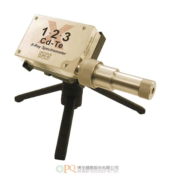
醫用 XR-100CdTe 探測器
Medical XR-100CdTe Detector 型號:X-123CdTe-
探測器系統設計用於同時測量 X 射線管峰值電位(kVp)和乳腺 X 射線管光譜圖。
產品特點- 直接測量光譜
- 終點能量(kVp)
- 檢視患者資料:
- 無康普頓光譜 ( Compton Spectra )
- 通過 XRS-FP 軟體進行逃逸峰調整
- 自我校正系統
- 直接對準 X 射線管,同時記錄光譜和峰值電位(kVp)
- 火星探路者技術現在也被用於放射學!
- 每個放射科必備的探測器
- 適用於射線照相和螢光透視系統的品質保證
所需設備- X-123CdTe 完整 X 射線和伽馬射線光譜儀
- EXVC 准直器套件
- XRS-FP 逃逸峰調整軟體
設計目標該探測器系統設計用於同時測量 X 射線管峰值電位(kVp)和乳腺 X 射線管光譜。
測量的意義- 射線管光譜和峰值電位(kVp)都是影響膠片螢幕和乳腺 X 射線攝影中圖像品質的重要參數。
- 在現代乳房攝影系統中自動選擇合適靶區/濾光片組合的過程可能會受到不當 kVp 的影響。
- 在傳統設備中,用戶依賴於中央實驗室校正,因此在使用過程中很難對儀器進行校正。
完整系統包括:
- 探測器 – X-123CdTe
- 准直器套件 – EXVC
- 逃逸峰調整軟體 – XRS-FP
輸出光譜示例
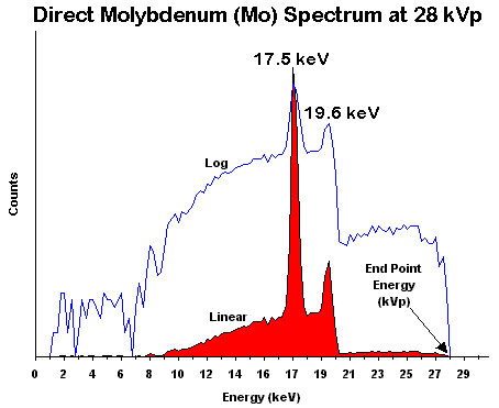
圖1. 乳房攝影機的 X 射線管監測
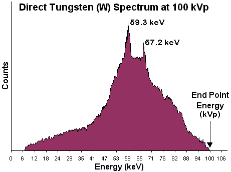
圖 2.放射機的 X 射線管監測
以上光譜由馬薩諸塞州伍斯特市馬薩諸塞大學醫學院 Andrew Karellas Ph.D 博士提供。 01655 USA
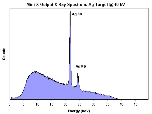
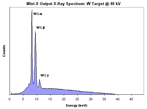
圖 3.Mini-X X 射線管的直接輸出 X 射線光譜(Ag 靶 – 頂部,W 靶 – 底部)
系統描述X-123CdTe
Amptek 的專長是 X 射線和伽馬射線光譜儀,它們輕巧、功耗低、性能高且操作簡單。 X-123CdTe 將 Amptek 的以下標準高性能 X 射線光譜測量元件整合在一個金屬盒中:XR-100CdTe 探測器和前置放大器、DP5 數位脈衝處理器和 MCA 以及 PC5 電源。 這就構成了一個可拿在手裡的完整系統。 在許多市售系統中,僅前置放大器的尺寸、品質和功率就比X-123CdTe的系統的體積還大。 它只需要 2 個介面即可運行:+5 VDC 電源和標準 RS-232、USB 或乙太網介面。 使用 X-123CdTe,任何人都可以快速獲得高品質的 X 射線和伽馬射線光譜。
准直器套件Amptek 開發了 EXVC 准直器套件,用於準直初級 X 射線束。 准直器金屬盒中最多可容納兩個鎢准直器片,這些准直器片被放置在探測器前面的接口支架內。 通過選擇適當的鎢准直器片,用戶可以減少入射的 X 射線通量,並允許探測器和電子設備處理 X 射線光譜。 Amptek提供了七種不同的鎢准直器片,它們有不同尺寸的孔(孔徑從 25 μm 到 2,000 μm 不等),適用於廣泛的應用。 准直器外殼由不鏽鋼製成。
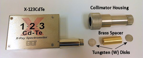
圖 4.EXVC 准直器套件
套件包括
- 不鏽鋼准直器外殼
- 三腳架和安裝板
- 鐳射筆
- 7 種鎢(W)准直器片:
- 1 mm 厚,帶 25 μm 孔
- 1 mm 厚,帶 50 μm 孔
- 2 mm 厚,帶 100 μm 孔
- 2 mm 厚,帶 200 μm 孔
- 2 mm 厚,帶 400 μm 孔
- 2 mm 厚,帶 1000 μm 孔
- 2 mm 厚,帶 2000 μm 孔
所有鎢片均由 HD17 合金(90% W、6% Ni、4% Cu)製成。所有鎢片和間隔管的直徑均為0.625英寸。
可選:EXVC-W-SPACER。此鎢(W)間隔管/準直器厚 36 mm,有 300 μm 的孔。 它設計用於阻止和準直由高能量射線管產生的大於 100 keV 的 X 射線。
使用 XRS-FP 軟體進行逃逸峰調整XRS-FP 軟體可用於調整 CdTe 探測器逃逸峰的輸出光譜。 這可能會對在 Cd 和 Te 吸收限上方運行的高能量 X 射線管產生顯著的效果。
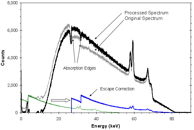
圖 5.該圖顯示了使用 CdTe 探測器採集的鎢(W)X 射線管輸出光譜在去除逃逸事件後的譜圖。 灰色軌跡線為原始光譜。 綠色軌跡線表示原始光譜中的逃逸事件。 這些事件在計算正確的能量(通過添加逃逸的能量)之前會從原始光譜中扣除。 藍色軌跡線表示校正後的逃逸事件,它們將與灰色軌跡線相加。 深黑色軌跡線表示處理后的最終結果,各事件都在正確的通道中。
參考文獻
- A. Karellas, I. Sechopoulos, I. Levis, A. C. Huber, and J. A. Pantazis, “Measurement of the x-ray spectra and tube potential in mammographic units with a self-calibrating compact cadmium zinc telluride (CZT) detector,” Radiology 205(P), 301 (1997).
- Matsumoto, Massao, et al. “Direct measurement of mammographic x-ray spectra using CdZnTe detector,” Medical Physics 27 (7), July 2000. p. 1490.
- Vedantham, Srinivasan, et al. “Mammographic imaging with a small CCD-based digital cassette: Physical characteristics of a clinical system,” Medical Physics 27 (8), August 2000, p. 1832.
- Vedantham, Srinivasan, et al. “Full breast digital mammography with an amorphous silicon-based flat panel detector: Physical characteristics of a clinical prototype,” Medical Physics 27 (3), March 2000, p. 558.
- S. Miyajima, “Thin CdTe detector in diagnostic x-ray spectroscopy,” Medical Physics, Vol. 30 No. 5, May 2003.
- S. Miyajima, K. Imagawa, M. Matsumoto, “An alignment method for mammographic X-ray spectroscopy under clinical conditions,” The British Journal of Radiology, 75 (2002), 763-766. © 2002 The British Institute of Radiology.
Abstract: This paper describes an alignment method for mammographic X-ray spectroscopy under clinical conditions. A pinhole, a fluorescent screen, a laser device and the case for a detector are used for alignment of the focal spot, a collimator and a detector. The method determines the line between the focal spot and the point of interest in an X-ray field radiographically. The method allows alignment for both central axis and off-axis directions. - S. Aiello, et al. “FLUXEN portable equipment for direct X-ray spectra measurements,” Nuclear Instruments & Methods in Physics Research, A 518 (2004) 389-390.
- P. Baldelli, et al. “Development of a quasi-monochromatic source for mammography applications,” Nuclear Instruments & Methods in Physics Research, A 518 (2004) 386-388.
- S. Miyajima and K. Imagawa, “CdZnTe detector in mammographic x-ray spectroscopy,” Physics in Medicine and Biology, 47 (2002) 3959-3972.
- S. Stumbo, U. Bottigli, B. Golosio, and P. Oliva, “Direct analysis of molybdenum target generated x-ray spectra with a portable device,” Med. Phys. 31 (10), p. 2763-2770, October 2004.
Abstract: In routine applications, information about the photon flux of x-ray tubes is obtained from exposure measurements and cataloged spectra. This approach relies mainly on the assumption that the real spectrum is correctly approximated by the cataloged one, once the main characteristics of the tube such as voltage, target material, anode angle, and filters are taken account of. In practice, all this information is not always available. Moreover, x-ray tubes with the same characteristics may have different spectra. We describe an apparatus that should be useful for quality control in hospitals and for characterizing new radiographic systems. The apparatus analyzes the spectrum generated by an x-ray mammographic unit. It is based on a commercial CZT produced by AMPTEK Inc. and a set of tungsten collimator disks. The electronics of the CZT are modified so as to obtain a faster response. The signal is digitized using an analog to digital converter with a sampling frequency of up to 20 MHz. The whole signal produced by the x-ray tube is acquired and analyzed off-line in order to accurately recognize pile-up events and reconstruct the emitted spectrum. The energy resolution has been determined using a calibrated x-ray source. Spectra were validated by comparison of the HVL measured using an ionization chamber. © 2004 American Association of Physicists in Medicine. - K. Maeda, M Matsumoto, A. Taniguchi, “Compton-scattering measurement of diagnostic x-ray spectrum using high-resolution Schottky CdTe detector,” Med. Phys. 32 (6), p. 1542-1547, June 2005. Available online.
- U. Bottigli, B. Golosio, G. L. Masala, P. Oliva, S. Stumbo, P Delogu, M. E. Fantacci, L. Abbene, F. Fauci, G. Raso, “Comparison of two portable solid state detectors with and improved collimation and alignment device for mammographic x-ray spectroscopy,”Med. Phys. 33 (9), p.3469-3477, September 2006. Available online.
- L. Abbene et al., “X-ray spectroscopy and dosimetry with a portable CdTe device,” Nuclear Instruments and Methods in Physics Research A 571 (2007) p. 373-377.
- G. Gerardi et al., “Digital filtering and analysis for semiconductor X-ray detector data acquisition,” Nuclear Instruments and Methods in Physics Research A 571 (2007) p. 378-380.
- Miceli, A., Thierry, R. et al.,”Comparison of simulated and measured spectra of an industrial 450 kV X-ray tube,” Nuclear Instruments and Methods in Physics Research A, 580 (2007) p.123-126.
- Josilene C. Santos, Alessandra Tomal, Tânia A. Furquim, Agnes M. F. Fausto, Maria S. Nogueira, Paulo R. Costa, “Direct measurement of clinical mammographic x-ray spectra using a CdTe spectrometer,” Medical Physics, 26 May 2017. Available online.
-
This detector system was designed to simultaneously measure the X-Ray tube peak potential (kVp), and to characterize the mammographic X-Ray tube spectrum.
Features
- Direct Measurement Spectra
- End Point Energy (kVp)
- See what the patient gets:
- No Compton Spectra
- Escape Peak Adjustment via XRS-FP Software
- Self-Calibrating System
- Look straight at the X-Ray tube and record simultaneously both the spectrum and the peak potential (kVp)
- The technology that went to Mars on the Pathfinder Mission is now available to Radiology!
- A must detector for every Radiology Department
- For Quality Assurance in Radiographic and Fluoroscopic Systems
Equipment needed- X-123CdTe Complete X-ray and gamma ray Spectrometer
- EXVC Collimator Kit
- XRS-FP Escape Peak Adjustment Software
Design Objective
This detector system was designed to simultaneously measure the X-Ray tube peak potential (kVp), and to characterize the mammographic X-Ray tube spectrum.
Significance of the Measurement- Both the tube spectrum and the peak potential (kVp) are important parameters affecting the image quality in film-screen and digital mammography.
- Automatic selection of proper target/filter combination in modern mammography systems may be affected by improper kVp.
- In conventional devices, the user depends on central laboratory calibration and has no easy way to calibrate the instrument during use.
Complete System Includes:- Detector – X-123CdTe
- Collimator Kit – EXVC
- Escape Peak Adjustment Software – XRS-FP
Example Output SpectraFigure 1. X-Ray Tube Monitor for Mammography Machines
Figure 2. X-Ray Tube Monitor for Radiology Machines
Above Spectra Courtesy of Dr. Andrew Karellas Ph.D, University of Massachusetts Medical School, Worcester, MA. 01655 USA
Figure 3. Direct output x-ray spectra from the Mini-X x-ray tube (Ag target – top, W target – bottom).
System Description
X-123CdTe
Amptek’s specialty is X-ray and gamma-ray spectrometers, which are small, low power, high performance, and simple to operate. The X-123CdTe combines in a single package Amptek’s standard, high performance X-ray spectroscopy components: the XR-100CdTe detector and preamplifier, DP5 digital pulse processor and MCA, and PC5 power supply. The result is a complete integrated system which can fit in your hand. In many commercially available systems, the preamplifier alone has more size, mass, and power than this integrated system. It requires only 2 connections to run: +5 VDC power and a standard RS-232, USB, or Ethernet connection. With the X-123CdTe, anyone can rapidly obtain high quality X-ray and gamma-ray spectra.
Collimator Kit
Amptek has developed the EXVC Collimator Kit to collimate the primary X-ray beam. The Collimator Housing can accommodate up to two Tungsten collimator disks that are placed inside a bayonet holder in front of the detector. By selecting the appropriate Tungsten collimator disks, the user can reduce the incoming X-ray flux and allow the detector and electronics to process the X-ray spectrum. Seven different Tungsten collimator disks are provided with different size holes (ranging from 25 µm to 2,000 µm hole) in order to allow for a wide range of applications. The Collimator Housing is made out of stainless steel.Figure 4. EXVC Collimator Kit. It can slide over the 1.5 inch extender of the X-123.
The kit includes
- Stainless steel collimator housing
- Tripod and mounting plate
- Laser pointer
- Brass Spacer
- 5 Tungsten (W) Collimator disks:
- 1 mm thick with 25 µm hole
- 1 mm thick with 50 µm hole
- 2 mm thick with 400 µm hole
- 2 mm thick with 1000 µm hole
- 2 mm thick with 2000 µm hole
All Tungsten disks are made of alloy HD17 (90% W, 6% Ni, 4% Cu).
All Tungsten disks and spacers have a diameter of 0.625 inches.
Optional: EXVC-W-SPACER
This Tungsten (W) Spacer /Collimator is 36 mm thick with a 300 µm hole. It is designed to stop and collimate x-rays greater than 100 keV produced from high energy tubes.Escape Peak Adjustment with XRS-FP Software
The XRS-FP software can be used to adjust the output spectrum for the escape peaks of the CdTe detector. This can be a significant effect for higher energy x-ray tubes that operate above the absorption edges of Cd and Te.For more information please see the application note ANCDTE1: CdTe Measurement of X-Ray Tube Spectra: Escape Events.
Figure 5. Plot showing a tungsten (W) x-ray tube output spectrum taken with a CdTe detector after processing to remove escape events. The gray trace shows the original spectrum. The green trace illustrates the escape events in the original spectrum. These are subtracted from this original spectrum, then the correct energies are computed (by adding in the energy which escaped). The blue trace shows the corrected escape events, which are then summed with the gray trace. The dark black trace shows the final result of the processing with the events in their correct channels.
References
- A. Karellas, I. Sechopoulos, I. Levis, A. C. Huber, and J. A. Pantazis, “Measurement of the x-ray spectra and tube potential in mammographic units with a self-calibrating compact cadmium zinc telluride (CZT) detector,” Radiology 205(P), 301 (1997).
- Matsumoto, Massao, et al. “Direct measurement of mammographic x-ray spectra using CdZnTe detector,” Medical Physics 27 (7), July 2000. p. 1490.
- Vedantham, Srinivasan, et al. “Mammographic imaging with a small CCD-based digital cassette: Physical characteristics of a clinical system,” Medical Physics 27 (8), August 2000, p. 1832.
- Vedantham, Srinivasan, et al. “Full breast digital mammography with an amorphous silicon-based flat panel detector: Physical characteristics of a clinical prototype,” Medical Physics 27 (3), March 2000, p. 558.
- S. Miyajima, “Thin CdTe detector in diagnostic x-ray spectroscopy,” Medical Physics, Vol. 30 No. 5, May 2003.
- S. Miyajima, K. Imagawa, M. Matsumoto, “An alignment method for mammographic X-ray spectroscopy under clinical conditions,” The British Journal of Radiology, 75 (2002), 763-766. © 2002 The British Institute of Radiology.
Abstract: This paper describes an alignment method for mammographic X-ray spectroscopy under clinical conditions. A pinhole, a fluorescent screen, a laser device and the case for a detector are used for alignment of the focal spot, a collimator and a detector. The method determines the line between the focal spot and the point of interest in an X-ray field radiographically. The method allows alignment for both central axis and off-axis directions. - S. Aiello, et al. “FLUXEN portable equipment for direct X-ray spectra measurements,” Nuclear Instruments & Methods in Physics Research, A 518 (2004) 389-390.
- P. Baldelli, et al. “Development of a quasi-monochromatic source for mammography applications,” Nuclear Instruments & Methods in Physics Research, A 518 (2004) 386-388.
- S. Miyajima and K. Imagawa, “CdZnTe detector in mammographic x-ray spectroscopy,” Physics in Medicine and Biology, 47 (2002) 3959-3972.
- S. Stumbo, U. Bottigli, B. Golosio, and P. Oliva, “Direct analysis of molybdenum target generated x-ray spectra with a portable device,” Med. Phys. 31 (10), p. 2763-2770, October 2004.
Abstract: In routine applications, information about the photon flux of x-ray tubes is obtained from exposure measurements and cataloged spectra. This approach relies mainly on the assumption that the real spectrum is correctly approximated by the cataloged one, once the main characteristics of the tube such as voltage, target material, anode angle, and filters are taken account of. In practice, all this information is not always available. Moreover, x-ray tubes with the same characteristics may have different spectra. We describe an apparatus that should be useful for quality control in hospitals and for characterizing new radiographic systems. The apparatus analyzes the spectrum generated by an x-ray mammographic unit. It is based on a commercial CZT produced by AMPTEK Inc. and a set of tungsten collimator disks. The electronics of the CZT are modified so as to obtain a faster response. The signal is digitized using an analog to digital converter with a sampling frequency of up to 20 MHz. The whole signal produced by the x-ray tube is acquired and analyzed off-line in order to accurately recognize pile-up events and reconstruct the emitted spectrum. The energy resolution has been determined using a calibrated x-ray source. Spectra were validated by comparison of the HVL measured using an ionization chamber. © 2004 American Association of Physicists in Medicine. - K. Maeda, M Matsumoto, A. Taniguchi, “Compton-scattering measurement of diagnostic x-ray spectrum using high-resolution Schottky CdTe detector,” Med. Phys. 32 (6), p. 1542-1547, June 2005. Available online.
- U. Bottigli, B. Golosio, G. L. Masala, P. Oliva, S. Stumbo, P Delogu, M. E. Fantacci, L. Abbene, F. Fauci, G. Raso, “Comparison of two portable solid state detectors with and improved collimation and alignment device for mammographic x-ray spectroscopy,”Med. Phys. 33 (9), p.3469-3477, September 2006. Available online.
- L. Abbene et al., “X-ray spectroscopy and dosimetry with a portable CdTe device,” Nuclear Instruments and Methods in Physics Research A 571 (2007) p. 373-377.
- G. Gerardi et al., “Digital filtering and analysis for semiconductor X-ray detector data acquisition,” Nuclear Instruments and Methods in Physics Research A 571 (2007) p. 378-380.
- Miceli, A., Thierry, R. et al.,”Comparison of simulated and measured spectra of an industrial 450 kV X-ray tube,” Nuclear Instruments and Methods in Physics Research A, 580 (2007) p.123-126.
- Josilene C. Santos, Alessandra Tomal, Tânia A. Furquim, Agnes M. F. Fausto, Maria S. Nogueira, Paulo R. Costa, “Direct measurement of clinical mammographic x-ray spectra using a CdTe spectrometer,” Medical Physics, 26 May 2017. Available online.